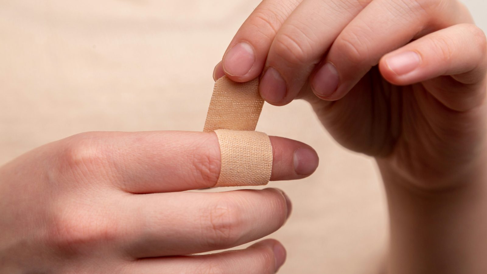
Skin healing is often described as though it were a simple process: damage occurs, new tissue grows and eventually the wound disappears. The biological reality is considerably more remarkable. Wound repair is a coordinated sequence involving platelets, immune cells, keratinocytes, fibroblasts, blood vessels, extracellular matrix and molecular signals operating across overlapping phases. Understanding that process changes the way we should think about claims that a botanical ingredient can heal skin.
When we cut a finger, graze a knee or damage the surface of the skin, the eventual disappearance of the injury can make healing seem almost effortless. It is anything but. Within seconds of tissue damage, blood vessels change their behaviour, platelets accumulate, coagulation pathways activate, immune cells begin arriving, chemical signals alter the behaviour of surrounding cells, fibroblasts migrate, new blood vessels develop, keratinocytes move across the wound surface, and collagen is deposited and reorganised. These processes do not occur independently. They communicate continuously, and what eventually appears to us as healed skin is the result of an extraordinarily coordinated biological response involving cellular behaviour, inflammation, vascular biology, structural proteins and biochemical signalling, much of it rebuilding the same barrier structures described in The Skin Barrier.
Wound healing is commonly divided into several overlapping phases: haemostasis, inflammation, proliferation and remodelling. The divisions are useful for understanding the process, but the body does not operate according to four neatly separated chapters. Inflammation begins while haemostasis is occurring, proliferation begins before inflammation has completely resolved, and remodelling begins while new tissue is still being constructed. Healing is better understood as a continuously changing biological environment, and that distinction becomes important when we encounter claims that an ingredient promotes wound healing. Before asking whether something accelerates healing, we need to understand what healing actually involves.
What exactly is a wound?
A wound occurs when the structural integrity of tissue is disrupted. At its simplest, this may involve damage limited largely to the epidermis, while more substantial injuries can extend through the dermis and into subcutaneous tissues, disrupting blood vessels, extracellular matrix, nerves and other structures. The biological challenge differs according to the severity of the injury. A superficial abrasion is not equivalent to a surgical incision, a surgical incision is not equivalent to a burn, and an acute traumatic wound is not equivalent to a chronic diabetic ulcer. This matters enormously when interpreting research, because an ingredient that appears to influence healing in an experimental incision in a healthy laboratory animal cannot automatically be assumed to improve healing in a person with a chronic diabetic foot ulcer. The biological environments are profoundly different.
Healing begins within seconds
The body cannot wait several hours before responding to an injury. The first priority is immediate: stop the bleeding. Damage to blood vessels exposes structures that normally remain hidden from circulating blood, platelets recognise the damaged environment and become activated, adhering to the site of injury, aggregating with one another and participating in formation of the initial haemostatic plug. At the same time, the coagulation cascade generates fibrin, which forms a network that stabilises the developing clot. This structure is more than an emergency patch. The provisional matrix created within the wound also provides a temporary environment through which cells can subsequently migrate. Repair has already begun.
Phase one: haemostasis
Haemostasis is sometimes omitted from simplified descriptions of wound healing and incorporated into the inflammatory phase, but it deserves separate attention. Immediately after vascular injury, local vasoconstriction helps reduce blood loss, platelets adhere and aggregate, and coagulation produces fibrin, forming a clot. Platelets are not merely passive building materials, however. Activated platelets release signalling molecules and growth factors that influence subsequent stages of repair, including mediators capable of communicating with fibroblasts, immune cells and vascular cells. The clot therefore performs two jobs simultaneously: it provides mechanical haemostasis, and it becomes part of the biochemical signalling environment that initiates repair. Within a remarkably short period, the wound has already begun transitioning into inflammation.
Phase two: inflammation
The word inflammation has acquired an overwhelmingly negative reputation in wellness and skincare. Products promise to reduce it, diets claim to eliminate it, and botanical ingredients are frequently celebrated for being anti-inflammatory. Yet without inflammation, normal wound healing would be impossible. Inflammation is not simply damage. It is an organised biological response to damage. Following injury, immune cells enter the wound environment, with neutrophils among the earliest leukocytes to arrive, participating in removal of microorganisms and damaged material and releasing numerous signalling molecules. Macrophages subsequently become particularly important, removing cellular debris, participating in microbial defence, releasing cytokines and growth factors, and critically, changing their behaviour as the wound environment evolves. Early inflammatory macrophage activity assists defence and clearance, while later macrophage phenotypes participate in resolution of inflammation and promotion of tissue reconstruction.
This transition is essential. A wound needs inflammation, but it also needs inflammation to resolve. Too little inflammatory activity can impair defence, while persistent or dysregulated inflammation can prevent the wound from progressing normally into repair. The objective of healthy healing is therefore not simply to suppress inflammation. It is to regulate it appropriately, and that distinction becomes extremely important when evaluating botanicals described as anti-inflammatory.
Phase three: proliferation
Once the initial inflammatory environment begins to change, the wound increasingly becomes a construction site. The proliferative phase involves several processes occurring simultaneously: fibroblasts migrate and proliferate, extracellular matrix is deposited, new blood vessels develop, keratinocytes move across the wound surface, granulation tissue fills the defect, and the wound begins contracting. These processes collectively transform a temporary inflammatory environment into new tissue, and scientific reviews describe the proliferative phase as involving fibroplasia, re-epithelialisation, angiogenesis and extracellular-matrix deposition. It is here that many experimental botanical studies report potentially interesting effects, but each process needs to be considered separately.
Granulation tissue
Healthy granulation tissue has a characteristic pink or red appearance because it contains numerous newly developing blood vessels, and is rich in fibroblasts, extracellular matrix, immune cells and vascular structures, effectively beginning to fill the space created by injury. Fibroblasts play a central role, migrating into the wound and producing components of the extracellular matrix that provide structural support for developing tissue, and some differentiate into myofibroblasts, cells possessing contractile properties that contribute to drawing the wound margins towards one another in a process known as wound contraction, which can substantially reduce the area that ultimately requires re-epithelialisation. When an animal experiment reports increased granulation tissue following treatment with a botanical extract, this represents an interesting biological observation, but it does not, by itself, establish that the botanical improves clinical wound healing in humans.
Angiogenesis
New tissue requires oxygen and nutrients, and existing circulation has often been damaged by the original injury, so the wound needs new blood vessels. Angiogenesis describes the development of new vascular structures from existing vessels, as endothelial cells respond to signals within the wound environment and participate in forming new capillary networks that supply the metabolically active tissue being constructed. The abundance of newly developing vessels is one reason healthy granulation tissue appears red, and as healing progresses and metabolic demand changes, many of these temporary vascular structures subsequently regress. The wound is continually being built and dismantled at the same time.
Re-epithelialisation
While deeper tissue is being reconstructed, something equally remarkable occurs at the surface. Keratinocytes, the predominant cells of the epidermis, begin migrating from the wound edges and surviving structures such as hair follicles, and their behaviour changes: rather than simply maintaining their normal position within the epidermis, they become migratory, moving across the provisional wound surface, proliferating, and communicating with fibroblasts and immune cells. Eventually, advancing epithelial fronts meet and restore continuity across the wound in a process called re-epithelialisation, one of the most commonly measured outcomes in experimental wound-healing research. Faster re-epithelialisation can be biologically meaningful, but again context matters, since an ingredient capable of altering keratinocyte migration in a cell culture does not automatically accelerate clinically meaningful wound closure in a person. The journey from Petri dish to patient is enormous.
Collagen and the extracellular matrix
Few biological molecules have become as commercially famous as collagen. In wound healing, however, collagen is not a beauty ingredient. It is structural architecture. Fibroblasts produce collagen and other extracellular-matrix components as new tissue develops, and early wound tissue contains substantial amounts of type III collagen; as healing progresses, the extracellular matrix is remodelled and relatively more type I collagen becomes incorporated, a transition that contributes to increasing mechanical strength. But the process is not simply about producing as much collagen as possible. Collagen fibres must be deposited, organised, cross-linked, degraded, replaced and reorganised, and an experiment showing increased collagen production should not automatically be interpreted as showing better healing, since excessive collagen deposition can itself be pathological. Healthy healing depends upon regulated matrix production and remodelling.
Phase four: remodelling
A wound can appear closed long before healing is complete, and remodelling may continue for months. During this period, extracellular matrix is repeatedly reorganised, collagen composition changes, fibres become more organised, unnecessary vascular structures regress, cellularity decreases, and tensile strength gradually increases. Yet repaired tissue does not normally return perfectly to its original state. Scar tissue differs structurally from unwounded skin, hair follicles, sweat glands and the intricate architecture of normal dermis may not be completely restored following deeper injury, and even mature scar tissue generally does not regain the full mechanical strength of uninjured skin. Healing is therefore not biological time travel. The body does not simply recreate the tissue that existed before injury. It repairs the defect as effectively as circumstances permit.
Why wounds sometimes fail to heal
The sequence described above assumes relatively normal physiology, and many wounds do not encounter such favourable circumstances. Healing can be disrupted by inadequate blood supply, infection, diabetes, repeated pressure or trauma, foreign material, poor oxygenation, nutritional deficiencies, immune dysfunction, certain medicines, smoking, advanced age, substantial tissue loss and persistent inflammation. Chronic wounds can become trapped within abnormal inflammatory environments, and instead of moving successfully through inflammation into proliferation and remodelling, the biological process stalls: proteases may remain excessively active, extracellular matrix can be degraded, cellular responses may become impaired, and microbial biofilms can further complicate the wound environment.
This is one reason simplistic statements such as calendula heals wounds are scientifically unsatisfactory. Which wound, in whom, at what stage, using which preparation, at what concentration, compared with what treatment, measured by which outcome? Those questions transform a marketing claim into a scientific question.
What scientists mean by wound-healing activity
The phrase appears frequently in botanical research, and it can mean many different things. Researchers may measure wound area, time to wound closure, re-epithelialisation, fibroblast proliferation, fibroblast migration, keratinocyte migration, collagen deposition, hydroxyproline concentration, granulation tissue, angiogenesis, inflammatory cytokines, oxidative stress, tensile strength or microbial burden. These are not interchangeable outcomes. An extract might alter inflammatory signalling without changing wound closure, another might stimulate fibroblast proliferation in vitro but fail to produce a clinically useful effect, and a third may appear to accelerate contraction in rodents, whose skin heals differently from human skin. Consequently, saying that an ingredient has demonstrated wound-healing activity without explaining what was actually measured tells the reader surprisingly little.
Why laboratory evidence is not clinical evidence
Botanical research becomes much easier to interpret if we imagine an evidence ladder. At the bottom are chemical and biochemical observations: a plant contains particular molecules, which is interesting but not evidence that it heals wounds. Next come in-vitro experiments, where an extract changes the behaviour of cultured fibroblasts, keratinocytes or inflammatory cells, giving us biological activity but still not clinical efficacy. Then come animal models, where a treated wound may close more quickly or demonstrate altered collagen deposition, stronger evidence that the effect can occur within living tissue, though animal skin is not human skin. Next come human observational and pilot studies, which begin to tell us whether an intervention is feasible and potentially useful, and finally appropriately designed randomised controlled clinical trials, systematic reviews and replicated evidence allow substantially greater confidence. The higher we climb, the more difficult and expensive the research becomes, and unfortunately many botanical claims climb the ladder much faster than the evidence does. This is the same discipline set out in our own Editorial and Evidence Policy.
Calendula and wound healing
Few botanicals are more closely associated historically with damaged skin than Calendula officinalis, examined in full in our Calendula monograph and in Can Calendula Really Heal Skin?. Calendula flowers have been incorporated into ointments, oils and other preparations for generations, and modern phytochemical research has identified numerous constituents, including triterpenoids, flavonoids, carotenoids and other compounds with potentially relevant biological activities. Of particular interest are triterpenoid compounds; research into triterpenes more broadly has identified effects involving cell migration, proliferation, oxidative processes and collagen deposition in experimental wound models, providing a plausible mechanistic basis for investigating calendula. But plausibility is not proof.
A systematic review published in Wound Repair and Regeneration evaluated animal and human studies of calendula extract, and across several animal studies, calendula treatment was associated with faster resolution of the inflammatory phase and increased granulation tissue, suggesting calendula extracts may influence biological processes occurring during the transition from inflammation into tissue reconstruction. The same review, however, demonstrates exactly why caution is necessary, since human results were inconsistent: some studies involving venous ulcers reported reductions in wound area, a trial involving diabetic leg ulcers did not demonstrate improvement, and burn studies and trials investigating radiation dermatitis also produced mixed and inconsistent findings. The biological signal is therefore more convincing than the clinical conclusion.
The European Medicines Agency provides an unusually useful illustration of how this evidence should be interpreted. Its Committee on Herbal Medicinal Products recognises calendula flower preparations as traditional herbal medicinal products for minor skin inflammation and as an aid in healing minor wounds, which sounds impressive until we examine what the classification actually means. It is specifically a traditional-use conclusion, not a finding of well-established clinical efficacy, and the EMA states that clinical evidence is insufficient to draw firm conclusions and that the recognised use rests upon longstanding use and biological plausibility. Calendula has not been dismissed, nor has it been clinically proven to be an effective general wound-healing treatment. The responsible conclusion lies between those positions: a scientifically plausible traditional botanical with encouraging preclinical evidence and insufficient clinical evidence for strong therapeutic wound-healing claims. That is considerably less exciting than calendula heals wounds. It is also considerably more accurate.
Lavender and wound healing
Lavender presents a different scientific story. Lavandula angustifolia essential oil contains a complex mixture of volatile compounds, with linalool and linalyl acetate among its major constituents, examined further in our Lavender monograph. Laboratory research has identified antimicrobial and anti-inflammatory activities relevant to wound biology, and perhaps the most frequently discussed experimental wound study was published by Mori and colleagues in 2016, who created full-thickness wounds in rats and applied lavender oil topically. Compared with controls, lavender-treated wounds showed reduced wound area during several time points, and the researchers also reported increased numbers of fibroblasts and altered expression of type I and type III collagen. Importantly, they investigated a potential mechanism involving transforming growth factor beta, an important signalling molecule within wound repair that influences fibroblast activity and extracellular-matrix deposition. The study therefore went beyond simply observing that wounds became smaller, attempting to identify a molecular pathway through which the effect might occur, which makes it scientifically interesting. But it remains a rat study.
A later review examining lavender essential oil and wound healing identified human trials, animal studies and in-vitro work, and taken collectively, the literature suggested potentially faster healing, altered collagen expression and effects upon proteins involved in tissue remodelling. However, the authors also identified major limitations. Lavender essential oil is not a single chemically identical substance; its composition varies according to species, cultivar, geography, harvesting, distillation and storage, so different studies may effectively be testing different chemical mixtures. Clinical studies are also heterogeneous, involving different wounds, different preparations, different concentrations, different comparator treatments and different outcomes, and the review’s authors called for larger, more standardised trials before lavender oil could be recommended as a validated clinical wound-care treatment. As with calendula, an intriguing rat model and a scientifically named mechanism do not, on their own, constitute proof of a clinical effect in people.
What the evidence allows us to say
Taken together, the calendula and lavender literature illustrate the same underlying lesson from two different angles. Both botanicals possess genuine, chemically identifiable constituents capable of influencing processes relevant to wound repair, including inflammatory signalling, fibroblast behaviour and collagen deposition. Both have produced encouraging findings in cell culture and animal experiments. And in both cases, the human clinical evidence is smaller, more heterogeneous and less conclusive than the preclinical evidence, for reasons that recur throughout botanical science: preparations differ, concentrations differ, wound types differ, and study quality varies considerably. Neither calendula nor lavender should be dismissed as inactive. Neither should be described as a proven clinical treatment for wounds. The accurate position, for both, sits between those two extremes, and it is the position this Library tries to hold consistently across every ingredient we examine.
What skincare can reasonably do
None of this means botanical skincare has no place in ordinary skin care. A cosmetic product does not need to heal a wound to be useful. Formulations built around calendula, lavender or similar botanicals can reasonably support skin comfort, provide emollience, contribute antioxidant or mildly anti-inflammatory constituents, and form part of a considered skincare routine for skin that is intact, dry or mildly irritated, exactly the territory explored in our calendula evidence article. What such a product should not claim is that it heals wounds, treats disease, or substitutes for medical care, because the clinical evidence for those specific claims does not yet exist at the standard that would justify them.
When botanical skincare is inappropriate
There are situations in which botanical skincare, however well formulated, is simply the wrong tool. Deep, infected, heavily bleeding or non-healing wounds require medical assessment rather than a cosmetic preparation. Chronic wounds associated with diabetes, impaired circulation or pressure require specialist wound care, compression therapy or other clinically directed treatment. Burns beyond the most minor and superficial require appropriate medical attention. And anyone using a botanical product on genuinely damaged skin should understand that a cosmetic formulated for intact skin is not automatically safe or appropriate for an open wound. Recognising the limits of what a jar of calendula or lavender cream can do is not a criticism of either botanical. It is simply an honest description of what cosmetic products are, and are not, designed to achieve.
References
- Givol, O. et al. (2019). A systematic review of Calendula officinalis extract for wound healing. Wound Repair and Regeneration, 27(5), 548-561. doi:10.1111/wrr.12737
- European Medicines Agency, Committee on Herbal Medicinal Products (HMPC). European Union herbal monograph on Calendula officinalis L., flos, revision 1. EMA/HMPC/437450/2017, adopted 27 March 2018. EMA/HMPC monograph
- Mori, H-M., Kawanami, H., Kawahata, H. & Aoki, M. (2016). Wound healing potential of lavender oil by acceleration of granulation and wound contraction through induction of TGF-β in a rat model. BMC Complementary and Alternative Medicine, 16, 144. doi:10.1186/s12906-016-1128-7
- Samuelson, R., Lobl, M., Higgins, S., Clarey, D. & Wysong, A. (2020). The effects of lavender essential oil on wound healing: a review of the current evidence. Journal of Alternative and Complementary Medicine, 26(8), 680-690. doi:10.1089/acm.2019.0286
Continue exploring
Botanical Library · Monograph No. 005
Calendula, Calendula officinalis
The complete ingredient monograph, including a dedicated evidence-for-skin section covering wound care, radiation dermatitis and venous ulcers.
Journal · Skincare
Can Calendula Really Heal Skin?
The accessible companion piece applying this wound-biology framework specifically to calendula’s evidence base.
Botanical Library · Monograph No. 001
Lavender, Lavandula angustifolia
The complete Lavender monograph, including its chemistry, traditional uses and cosmetic applications.